Download here the brochure CenterVue EIDON AF | Confocale scanner.
0%


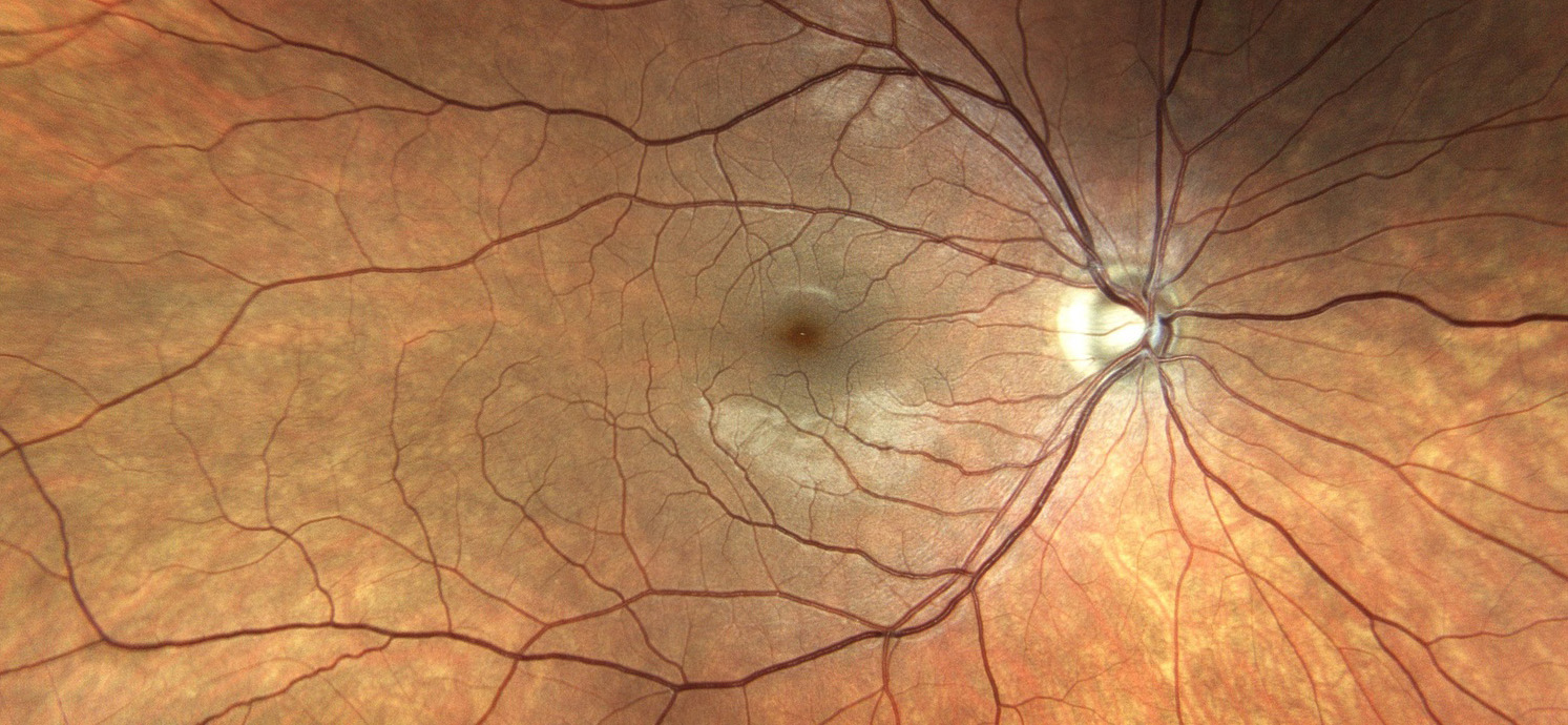

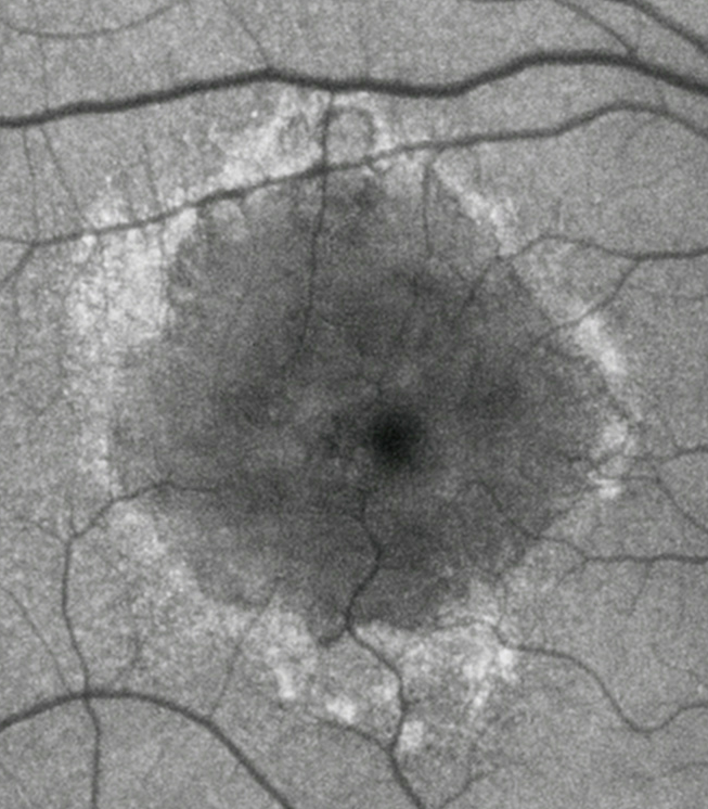


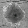
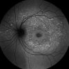
Fundus Autofluorescence (FAF) imaging is a non-invasive technique which provides information on retinal metabolism and health, through showing changes in the integrity of the Retinal Pigment Epithelial (RPE) layer. FAF imaging may help to understand metabolic alterations of the RPE in the pathogenesis of several retinal disorders. FAF provides these clinically useful information by taking an image based on the distribution pattern of fluorescent pigments accumulating in the RPE, such as lipofuscin. RPE dysfunction is visualized as an increased FAF signal, that is bright areas in the image, corresponding to lipofuscin accumulation RPE or photoreceptor death is visualized as a decreased FAF signal. That is dark areas in the image, corresponding to lipofuscin absence.
Download here the brochure CenterVue EIDON AF | Confocale scanner.
![]()
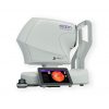
Eidon AF represents the natural evolution of Eidon TrueColor Confocal Scanner, including all its features and functionalities, preserving its unsurpassed quality of image, and adding autofluorescence imaging capabilities.
Eidon AF with Autofluorescence capability is now an extraordinary tool for obtaining multiple types of highvalue information from multiple imaging modalities:
| Dr. SriniVas R. Sadda introduces CenterVue Eidon AF and highlights the importance of Autofluorescence in clinical practice. |
Je moet inloggen om een reactie te kunnen plaatsen.
Reviews
There are no reviews yet, would you like to submit yours?